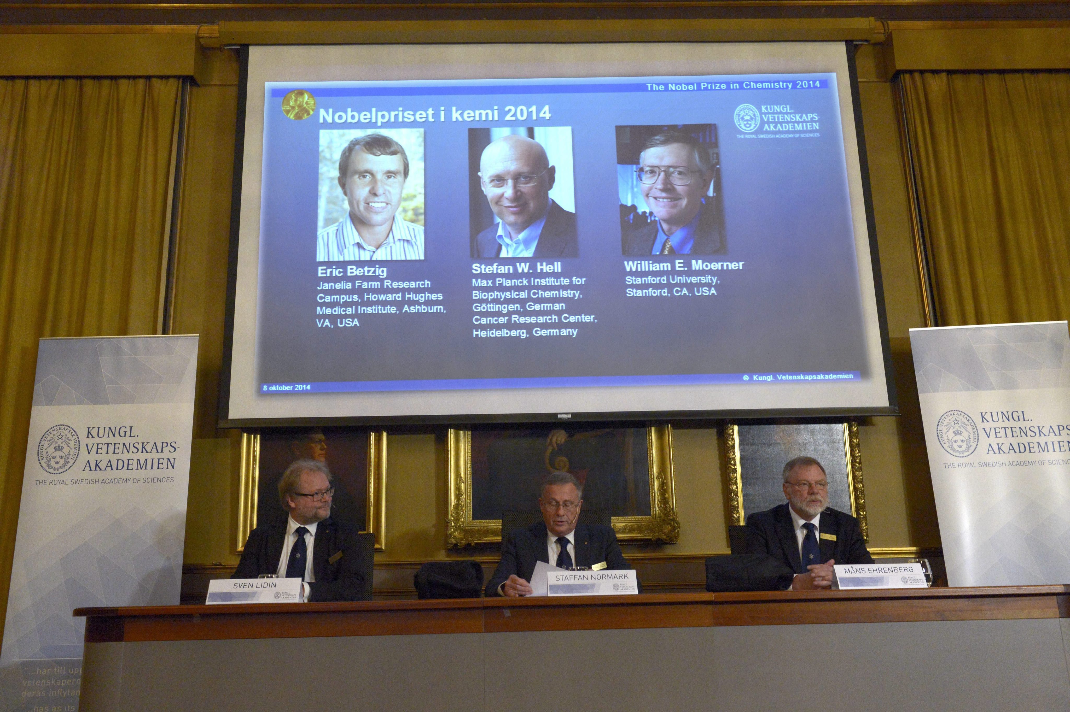
A Real Academia Sueca das Ciências reconheceu-lhes o trabalho e atribuiu-lhes esta quarta-feira o Nobel da Química de 2014, justamente por terem, trabalhando em separado, conseguido aumentar a capacidade dos microscópios a uma resolução maior do que “metade do comprimento de onda da luz”, segundo se lê no comunicado da instituição.
Durante anos pensou-se que esta era a medida do limite dos microscópios. O que os premiados introduziram de novo foi a capacidade de visualizar “o trajecto de moléculas individuais dentro de células vivas. Eles podem ver de que modo as moléculas criam sinapses entre células nervosas no cérebro; podem localizar proteínas envolvidas nas doenças de Parkinson, de Alzheimer e de Huntington”, entre outras barreiras que se quebraram.
O processo ficou conhecido como nanoscopia e abriu caminho a potentes observações, a locais onde a ciência ainda não tinha chegado, e à possibilidade de uma nova compreensão de processos biológicos e doenças e a novas terapias.
O prémio será entregue em Estocolmo, a 10 de Dezembro, e os três vencedores irão repartir um total de 867 mil euros.

O anúncio do nomes dos três vencedores do prémio Nobel Bertil Ericson/EPA
As explicaçoes da Aseembleia do Nobel:
"In what has become known as nanoscopy, scientists visualise the pathways of individual molecules inside living cells. They can see how molecules create synapses between nerve cells in the brain; they can track proteins involved in Parkinson’s, Alzheimer’s and Huntington’s diseases as they aggregate; they follow individual proteins in fertilised eggs as these divide into embryos.
It was all but obvious that scientists should ever be able to study living cells in the tiniest molecular detail. In 1873, the microscopist Ernst Abbe stipulated a physical limit for the maximum resolution of traditional optical microscopy: it could never become better than 0.2 micrometres.
Eric Betzig, Stefan W Hell and William E Moerner are awarded the Nobel Prize in Chemistry 2014 for having bypassed this limit. Due to their achievements the optical microscope can now peer into the nanoworld.
Two separate principles are rewarded. One enables the methodstimulated emission depletion (STED) microscopy, developed by Stefan Hell in 2000. Two laser beams are utilised; one stimulates fluorescent molecules to glow, another cancels out all fluorescence except for that in a nanometre-sized volume. Scanning over the sample, nanometre for nanometre, yields an image with a resolution better than Abbe’s stipulated limit.
Eric Betzig and William Moerner, working separately, laid the foundation for the second method, single-molecule microscopy. The method relies upon the possibility to turn the fluorescence of individual molecules on and off. Scientists image the same area multiple times, letting just a few interspersed molecules glow each time. Superimposing these images yields a dense super-image resolved at the nanolevel. In 2006 Eric Betzig utilised this method for the first time.
Today, nanoscopy is used worldwide and new knowledge of greatest benefit to mankind is produced on a daily basis."


